| Return to Main UDAAN Page |
The hard disk of the computer you are using right now stores the information as 0 or 1, in sets of 8 such digits, making a code of 28 or 256 character ASCII code, that stores all information whether it is a software or data. Similarly, the cells in our body stores all genetic information as a double stranded long tape like chemical molecule, made out of 4 chemical bases (unlike 2 of the computer), arranged in sets of three (unlike 8 in the computer), to give the code for all the amino acids in the body, and other information. The bases are bonded together with the help of Deoxyribose sugar (hence the name Deoxyribose Nucleic Acid or DNA) and phosphate bonds.
|
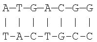
A very simplified DNA chain looks like this. The four bases are Adenine, Thymine, Cytosine and Guanine. The bases always combine in the form of A with T and C with G, as shown in the figure. The horizontal row is the strand of bases, and two such strands as shown are coiled into a double helix structure to make the genetic code. There are 46 chromosomes, arranged in the form of 23 pairs. |
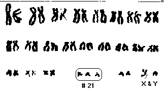
| The first 22 pairs of chromosomes are somewhat
similar and called autosomal, while the 23rd pair may be dissimilar and is
called sex chromosomes, since they determine sex. If they are similar,
i.e. X and X, then the person is a female. If they are not similar, i.e. X
and Y, as in this picture, then the person is a male.
Down Syndrome occurs when the 21st pair has an extra chromosome (Trisomy 21), as shown. Sometimes, the extra chromosome may join with another chromosome (14, 21 or 22) or get translocated. |
 |
In addition, the trisomy 21 may be present since inception, or take place as a mutation during cell growth, or some of the trisomy 21 cells may lose the third chromosome during cell growth. In the latter two cases, some cells have trisomy 21 while some have the normal pair only. This state is called mosaicism.
Out of the 23 pairs of chromosomes, the 21st pair of chromosomes are the smallest, and contain only about 250 or less genes. Out of them, some 2 to 4 dozen genes are such that if they are over-expressed due to triplication, they may produce the abnormalities of Down Syndrome. These genes are not at one place but scattered, and not all of them have been identified yet.
Due to trisomy 21, some genetic information gets over-expressed, contributing to the various developmental abnormalities associated with Down Syndrome. Some important defects suspected from the over-expression of genes due to trisomy 21 are given below. Please note that NO gene has been definitively linked to Down Syndrome, though the ones given below are strong contenders. Even within the same chromosome, the alleles may differ in their placement and expression from one person to another, hence Down Syndrome presents in a wide variety of features and marked inter-individual variation. Lastly, maternal age has a strong bearing on the development of Down Syndrome, as per statistics quoted later on.
The Suspected Defects |
Their possible culprit genes |
|
Superoxide Dismutase (SOD1) |
|
COL6A1 |
|
ETS2 |
|
CAF1A |
|
Cystathione Beta Synthase (CBS) |
|
DYRK |
|
CRYA1 |
|
GART |
|
IFNAR |
|
APP, GLUR5, S100B, TAM, PFKL Some others not yet identified |
| Age (years) | Frequency of
Fetuses with Down Syndrome to Normal Fetuses at 16 weeks of pregnancy |
Frequency of Live
Births of Babies with Down Syndrome to Normal Births |
|---|---|---|
|
15 - 19 |
---- |
1 / 1250 |
|
20 - 24 |
---- |
1 / 1400 |
|
25 - 29 |
---- |
1 / 1100 |
|
30 - 31 |
---- |
1 / 900 |
|
32 |
---- |
1 / 750 |
|
33 |
1 / 420 |
1 / 625 |
|
34 |
1 / 325 |
1 / 500 |
|
35 |
1 / 250 |
1 / 350 |
|
36 |
1 / 200 |
1 / 275 |
|
37 |
1 / 150 |
1 / 225 |
|
38 |
1 / 120 |
1 / 175 |
|
39 |
1 / 100 |
1 / 140 |
|
40 |
1 / 75 |
1 / 100 |
|
41 |
1 / 60 |
1 / 85 |
|
42 |
1 / 45 |
1 / 65 |
|
43 |
1 / 35 |
1 / 50 |
|
44 |
1 / 30 |
1 / 40 |
|
45 and older |
1 / 20 |
1 / 25 |
The zone written with yellow font is an increased risk zone while the zone written with red font is a high risk zone. The middle column above showing the occurrence rates at 16 weeks of pregnancy is mentioned because as per natural laws of survival of the fittest, many abnormal fetuses abort by this time.
Adapted from Hook EB. Journal of Am. Med. Assoc.; Vol. 249: Page 2034-2038, Year 1983.
| Return to Main UDAAN Page |