Summary: A brief bird's eye view of the
anatomical considerations behind various brain related disabilities, in lay
person's language.
Last updated January 21, 2007
Cerebral Palsy (CP) and Mental Retardation (MR) may be the
result of irreversible damage to one or more concerned areas of brain,
during intrauterine life, labor or infancy. A particularly vulnerable part
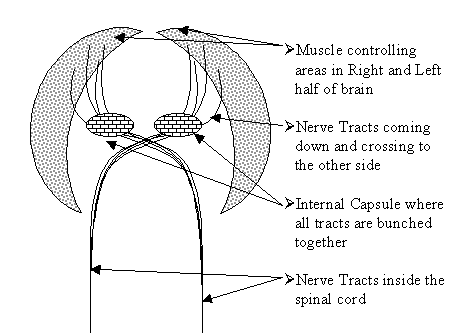
is the Internal Capsule region of brain where most nerve fibers from the
muscle controlling areas of brain come together to then pass down to the
spinal cord. This area has the poorest blood supply and is the most frequent
site of blood flow obstructive disorders, even in adult life.
Mental Retardation can also result from severe brain injury or infection
later on.
Even Autism is now linked to discrete areas of
brain damage, possibly caused by damage to the intracellular micro-tubules,
by incorporation of mercury into their walls, as a result of a genetic
defect in mercury excretion leading to its accumulation and intoxication.
Other causes like micro-nutrient deficiency, gut infections, psycho-social
causes, etc. only seem to be playing an aggravating role. SPECT Scan of some
our our Autistic / Autistic spectrum children have shown ischemic changes in
brain perfusion similar to CP.
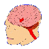
The nervous system central part is divided into the main brain (brown) and
the spinal cord. The brain is further subdivided into structural (anatomical)
parts and functional (physiological) parts. Structurally, the outer part front,
and sides and back top are called Cerebrum, while
the rear lower reddish brown part is called Cerebellum.
The outer layer is called cortex (containing most of the nerve cells or gray
matter) while the inner core is called white matter as it mostly contains nerve
tracts, and islands of nerve cell masses called nuclei.
Thus the full name of the thinking, motor and sensory part of the brain
is Cerebral Cortex, while the coordinating
part is called Cerebellar Cortex. On the
inside center are nuclei that control pain perception, involuntary
activity, and emotions, which together in turn control the hormonal
balance of the body.
Nerve fibers from the muscle controlling areas of brain come down and
bunch together at a place called Internal capsule. This area is notorious
for its poor blood supply and is almost always the victim of any
deficiency in blood supply, both in infants and adults. This leads to
paralysis of muscles controlled by those nerve fibers. In cerebral palsy
caused by low blood supply injury, this is a common site of injury.
Thus, in Cerebral (= main brain) Palsy
(= short form of paralysis), the affected person has reduced motor power,
reduced coordination of movement, and exaggerated tone (due to lack of
control over muscle tone generating power of the spinal cord, which tries
to contract the muscles, while the brain tries to relax them; the
interplay of these two forces controls the degree of muscle tone.)
The spinal cord motor nerve cells are like the accelerator of a car, always
trying to contract their corresponding muscle groups, while the cerebral Cortex
and cerebellum are like the brakes of a car, trying to control speed and power.
Hence, when the latter are malfunctioning, the body muscles become unduly
contracted or spastic, hence the paralysis or paresis (meaning incomplete
paralysis) is called Spastic paralysis.
A normal person with average intelligence is said to have an Intelligent
Quotient (IQ) of 100% for his age. A normal person may have an IQ of
about 100 + 10%. In Mental Retardation, a
person has an IQ significantly lower, often as low as 20 to 30% (severe cases)
or 40 to 60% (milder cases).
Regular guided exercises (physiotherapy),
relaxant drugs and injections of muscle relaxing poisons in micro doses
(poison extracted from the food poisoning bacteria, Clostridium botulinum,
called Botox or Botulinum Toxin), may
prolong the useful movement phase in the life of these cases. However, botulinum
toxin is of limited use as of today, since it gives only 2 to 4 months of relief
per course. Development of anti-toxin antibodies makes repeated courses not as
useful as the first one.
Hence, it is back to fun-filled physiotherapy and corrective (shortening or
lengthening surgical procedures on contracted tendons and muscles) operations as
and when required.
Contact
Person: Please help us avoid Virus attacks / Spam.
Ensure that your letter has a Subject line with a meaningful header
mentioning UDAAN otherwise our filter may send the letter to Trashcan.